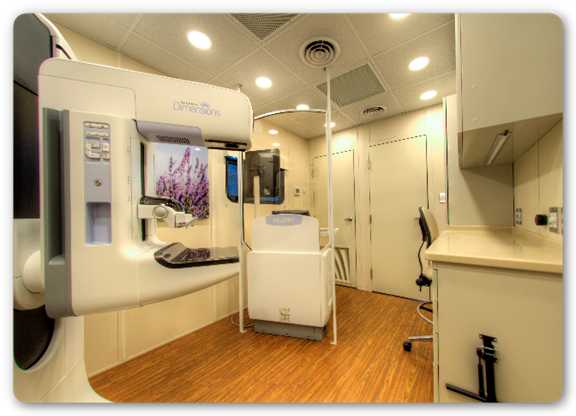
Breast tomosynthesis, known as three-dimensional (3D) mammography or digital breast tomosynthesis (DBT) is an advanced form of breast imaging that uses a low-dose x-ray system and computer reconstructions to create three-dimensional images of the breasts. This technology is far more advanced and accurate than typical 2D mammography and is just as safe.
A conventional mammogram provides a two-dimensional image of the breast utilizing two X-ray images of each breast. However, three-dimensional mammography (aka 3D tomosynthesis) uses several low dose images from different angles around the breast to create a 3D picture.
The data from a three year study on breast cancer mammogram screenings using 3D tomosynthesis identified significant benefits over time. This study, published February 18, 2016 by JAMA Oncology details the “Effectiveness of Digital Breast Tomosynthesis Compared with Digital Mammography: Outcomes Analysis from 3 Years of Breast Cancer Screening.”
The senior author of the study, Dr. Emily Conant, M.D., chief of breast imaging at the Perelman School of Medicine at the University of Pennsylvania and a highly respected member of the Breastcancer.org Professional Advisory Board, stated “These findings reaffirm that 3D mammography is a better mammogram for breast cancer screening.” Dr. Conant goes on to say the “results are an important step toward informing policies so that all women can receive 3D mammography for screening.”
According to experts at MD Anderson Cancer Center, women should consider 3D mammography breast screenings from either a fixed site, or from a roving mobile mammography unit. This FDA-approved 3D technology uses multiple breast images to create a 3D picture of the breast. These multiple images of breast tissue slices give doctors a clearer image of breast masses and makes it makes it much easier to detect breast cancer.
Mobile units with 3D tomosynthesis on board utilize higher voltage electrical power from a nearby building or an on-board power generator unit to power the 3D x-ray. Recently, a state-of-the-art 3D mobile unit rolled out by The James Cancer Center at Ohio State University’s Wexner Medical Center features a LifeLineMobile mammography unit with LifeLine’s unique U.S. patent pending power control system. This system enables The James Cancer Center to offer state-of-the-art 3D breast screening along with their professional expertise, continuity of care, convenience and support to patients throughout central Ohio.
Patients and their medical team find significant benefits from the 3D process because it develops multiple images of breast tissue to give doctors a clearer image of breast masses and more easily detects the presence of breast cancer. This unique 3D mammography is of no greater risk to the patient because it releases the same amount of low dose radiation as a traditional mammogram.
Conclusion
As advancements in mammography technologies continue and organizations mobilize into underserved populations, more and more lives are saved every day. Even the most advanced equipment cannot reach the thousands, or perhaps millions, of women in the over 40 year old age bracket in rural or urban communities if it is only available in a brick and mortar clinic. Though these facilities are vital to those in their communities, mobile mammography plays a critical role in encouraging advanced medical treatment to the underserved population.
Sources{{cta(‘138806f7-edd5-4f69-9291-dae2e0353da5′,’justifyright’)}}